Shape-shifting cell study explains why certain foods leave stomachs hungry
A study on rat brains reveals "brakes" in the brain that control satiety.

Digging into a meal is as much a psychological experience as it is a visceral one: As you shovel food down your throat, the brain prepares to tell you when it's time to stop. It's an instinctual process that's illuminated by new research: Now, scientists have a better idea of how and why the brain sends out that fullness signal, and why some kinds of foods might make it neglect that duty.
The feeling of fullness is governed by cells in the brain called astrocytes — star-shaped cells that outnumber neurons more than 5 to 1 in the brain. Scientists examined the brains of mice and found that when astrocytes are working properly, they change shapes. This study posits that shape-shifting allows neurons to send out a satiety signal, which ignites a feeling of fullness.
These results were published Tuesday in the journal Cell Reports.
Crucially, this study also shows that certain foods interfere with way the brain relays that fullness signal, whereas others might set it in motion.
According to this experiment, high-fat diets appear to affect how these astrocytes change their shape, in turn, suppressing satiety. But, in an unconventional twist sugar seemed to set the fullness signal in motion, explains lead study author Alexandre Benani, a researcher at the French National Centre for Scientific Research and the University of Burgundy in France.
"This work provides one neuronal basis that might explain the specific satiety index of nutrients, or at least, the difference between carbohydrates and lipids," he tells Inverse.
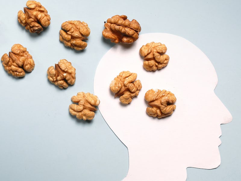
"In that way, sugar-rich foods such as potatoes have higher satietogenic properties than high-fat foods, such as nuts."
High calorie and high-fat diets may affect how the brain sends out a signal indicating fullness, according to a new mouse study.
Bad news for high-fat diets
This experiment dives into the minutia of the mouse brain to establish what exactly goes on during eating. To do this, the team looked at three groups of mice: one group that was given a standard diet ("balanced," as the authors put it), one that got a high-calorie and high-fat diet, and a third control group that ate nothing.
The team was able to zero in on the exact cells that are hyperactive during eating: the POMC neurons in the hypothalamus, an area deep in the brain that deals with the basic details of being alive, like reproducing or eating.
POMC neurons are thought to play a crucial role in the suppression of eating. They're more active when body temperature goes up (perhaps the reason eating feels unappealing when you're running a fever, or just finished a hard workout). When these cells are more active, we feel more full.
This team found that POMC neurons were three times more active in the group that ate their fill for one hour compared to the starved control group. But oddly, they found that the POMC neurons appeared less excitable in the mice who consumed a high-calorie, high-fat diet, compared to the mice who ate a balanced diet.
The study authors indicate that this finding suggests that whether or not those neurons get sufficiently activated depended on the kinds of foods that are eaten.
A "brake" on appetite
In the second part of the study, the team dug a bit deeper, this time, focusing on astrocytes – cells that wrap around POMC neurons in the brain. Past research has suggested that these cells act as "coverage" – like a brake, Benani explains. When that brake is engaged, they can insulate POMC cells, and keep their activity dampened, which allows one to keep eating without feeling full.
But if their shape is changed (imagine releasing a parking brake, for instance) they can allow the excitability of those cells to shine through. This experiment suggested that certain kinds of foods, especially high-fat or high-calorie foods, might affect whether or not those astrocytes "retracted" (or, to use the brake idea "released").
After a balanced meal, the team found that the astrocytes retracted as usual. But in response to the high-fat diet, the team found no significant change. They didn't retract, and in turn, blunted the activity of the POMC neurons.
As Benani explains, glucose seems to be one of the major factors that trigger that release. In an additional group of mice, he found that mice given a high-sugar diet also saw the activation of their POMC neurons —and those put on a high-fat diet did not experience the same level of activation.
"Astrocytes act as a brake applied on POMC neurons, and glucose causes astrocyte removal from POMC neurons, which ultimately increases the activity of these neurons," Benani says. "Note that we still do not know how astrocytes shift their shape: the underlying molecular mechanism is still unknown. We just show the crucial role of glucose as a triggering factor."
POMC neurons at the base of the mouse brain. These neurons inhibit eating, and perhaps signal fullness when astrocyte coverage retracts.
These results suggest that high-fat diets may make mice feel less full because they alter the way that astrocytes change their shape. When that coverage is never altered, it doesn't allow that brain signaling associated with satiety to shine through.
Still, the study was conducted in mouse brains — and research conducted on human brains is needed to say if this mechanism holds up in humans, Benani says.
"It's hard to directly transpose this result in humans, just because of the resolution of the scanners," he says. "However, the same brain structures are found in both murine and human brains, with the same POMC neurons and same functioning with anorectic POMC neurons."
This study gives us a micro-view of a bigger feeling: the process in the brain that drives that feeling of fullness. For now, it gives us something to visualize when the feeling of sleepy fullness hits. It's not increasing tightness of your waistband that's making you feel full, it's the shape-shifting cells in the brain.
Abstract: Mechanistic studies in rodents evidenced synaptic remodeling in neuronal circuits that control food intake. However, the physiological relevance of this process is not well defined. Here, we show that the firing activity of anorexigenic POMC neurons located in the hypothalamus is increased after a standard meal. Postprandial hyperactivity of POMC neurons relies on synaptic plasticity that engages pre-synaptic mechanisms, which does not involve structural remodeling of synapses but retraction of glial coverage. These functional and morphological neuroglial changes are triggered by postprandial hyperglycemia. Chemogenetically induced glial retraction on POMC neurons is sufficient to increase POMC activity and modify meal patterns. These findings indicate that synaptic plasticity within the melanocortin system happens at the timescale of meals and likely contributes to short-term control of food intake. Interestingly, these effects are lost with a high-fat meal, suggesting that neuroglial plasticity of POMC neurons is involved in the satietogenic properties of foods.