EVALI: 3 shocking images reveal the damage vaping can do to teen lungs
The lungs of 14 teenagers provide solid evidence of the damage e-cigarettes can do to health.

Teenagers still very much want to vape, despite the FDA’s attempt to make e-cigarettes look lame and researchers' efforts to prove vaping can harm the body. But a cache of new images might be enough to put some off their favorite habit.
Vaping is on the rise: An estimated 5.4 million American middle and high school students vape. Scientists are racing to become better at diagnosing EVALI, the lung injury illness associated with vaping.
In a new study, scientists reveal the damage the condition does to teen lungs for the first time. The study uses chest X-rays and computed tomography (CT) imaging to show patterns of EVALI-induced damage in these young people's lungs — the likes of which have only been seen in adults so far. But fifteen percent of reported EVALI cases are in people under the age of 18.
The new analysis was published Tuesday in the journal Radiology. It is the first of its kind.
What the scans show
This was a retrospective study of 14 adolescents, aged between 13 to 18, who were hospitalized for EVALI. The teens, 7 male and 7 female, underwent chest radiography and CT within 4 days of symptom presentation. All of the study participants either used an e-cigarette or “dabbed” — meaning they inhaled small quantities of a vaporized drug, like cannabis oil. Nine of them only used vapes containing TCH, four used THC and nicotine-containing vapes, while one person only used nicotine-containing vapes.
Two pediatric radiologists independently reviewed their scans for pattern, distribution, and extent of pulmonary and extrapulmonary abnormalities.
Reviewing X-rays and other chest scans is one of the most-accurate ways to diagnose EVALI. Symptom-wise, it is easy to confuse it with the flu and people can be reluctant to tell their doctors they vape. In this new study, all 14 of the subjects had respiratory symptoms, including shortness of breath, cough, and chest pain, and 12 of the subjects experienced gastrointestinal symptoms, too, including vomiting and diarrhea.
The key image finding was ground-glass opacity in the lungs. Ground-glass opacity is a type of lung damage that can be caused by the partial filling of air spaces, increased capillary blood volume, the partial collapse of alveoli (the tiny air sacs of the lungs).
The CT scans, and not the chest X-rays, also revealed that 36 percent of the participants with EVALI had the “reversed halo sign” — an imaging descriptor that has been reported in association with a range of other pulmonary diseases, including pneumonia and tuberculosis.
Seventy-nine percent of the patients' CT scans also showed signs of subpleural sparing. The subpleural regions of the lungs are found between the body wall and the membranes that envelop the lungs, as well as line the thorax. Subpleural sparing is when these regions are still preserved, despite damage elsewhere.
Here are three of the cases in the study, and what the radiologists saw:
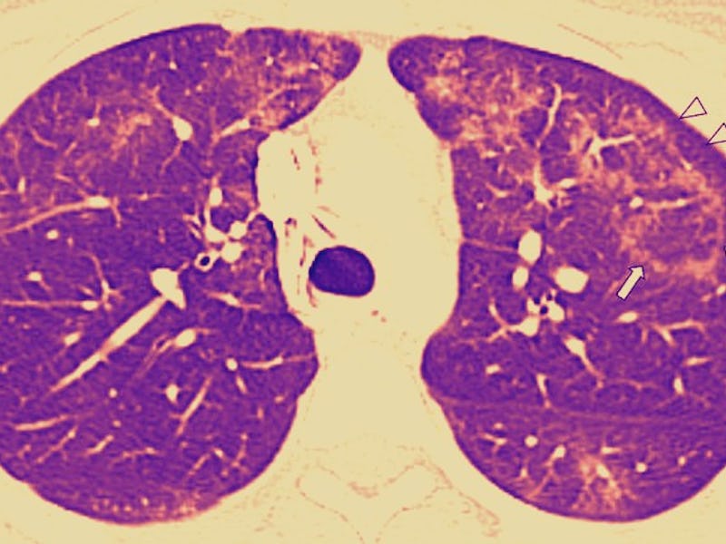
A chest CT scan.
This is a scan of the axial lung window. It gives radiologists a look at the middle regions of the lungs. These lungs belong to a 16-year-old girl who, for one year, used both nicotine and THC-containing e-cigarettes, until she experienced acute abdominal pain. After a day of vomiting, she went to the hospital. The white triangles in the image point to ground-glass opacity, surrounded by the reversed halo sign. The arrows also point to subpleural sparing — a sign of lung abnormalities.
An axial lung window image of CT with contrast enhancement performed.
This CT scan of an axial lung window belongs to a 17-year-old boy. He used THC-containing e-cigarettes for two months until he experienced 8 days of nausea, vomiting, abdominal pain, and fever. Those symptoms were paired with subsequent chest pain and shortness of breath. The arrows point to the presence of central ground-glass opacity, surrounded by the reversed halo sign.
A digital chest radiograph and a chest CT.
On the left, we have a posteroanterior digital chest radiograph taken the day the person came into the hospital, and on the right is a non-contrast chest CT taken the next day. The study participant in question is a 16-year-old with a 2-year history of daily vaping — both of nicotine, and THC. He went to the hospital after 1.5 weeks of coughing, shortness of breath, nausea, and vomiting. Both images show signs of ground-glass opacities.
What causes EVALI?
E-cigarettes can contain a variety of chemical constituents that can harm the lungs. But it is vitamin E acetate that has emerged as the leading cause of EVALI. It is typically used to thicken THC oil and, in turn, is often found within black market vapes. An analysis published in The New England Journal of Medicine determined that vitamin E acetate was present in 48 out of 51 lung tissue samples from people with EVALI.
The data suggest that, when vapor containing vitamin E acetate is inhaled, acute lung injury may occur. The authors of this paper also point out that the inhalation of e-cigarette aerosols can cause lung damage, too. They write that fibrous exudates within the alveoli could explain the presence of the ground-glass opacities, and generally signal the presence of substantial lung damage.
More studies are needed to say for sure — studies with more patients, and studies that specifically look at isolated cases of nicotine and THC vape use.
But these images make it clear that e-cigarettes can change and damage the lungs, and to get the full picture, it's best to combine CT scanning and X-rays.
Partial abstract:
Purpose: To evaluate chest radiographic and chest CT findings of EVALI in the pediatric population.
Results: Seven male patients (50%) and seven female patients (50%) (mean age, 16 years; range, 13–18 years) were evaluated. All patients underwent chest radiography and CT within 4 days of presentation (range, 0–4 days). Chest radiographic findings included ground-glass opacity in 14 of 14 (100%) and consolidation in eight of 14 (57%). CT findings included ground-glass opacity in 14 of 14 (100%), consolidation in nine of 14 (64%), and interlobular septal thickening in two of 14 (14%). At CT, subpleural sparing was seen in 11 of 14 (79%) and a reversed halo sign was seen in five of 14 (36%). Chest radiographic and CT abnormalities were predominately bilateral in 14 of 14 (100%) and symmetric in 13 of 14 (93%), with lower lobe predominance in seven of 14 (50%). Extent of abnormality was predominately diffuse at both chest radiography and CT. There was almost perfect interobserver agreement between two reviewers for detecting abnormalities on chest radiographs (k = 0.99; 95% confidence interval: 0.97, 1.00) and CT (k = 0.99; 95% confidence interval: 0.98, 1.00).
Conclusion: Conclusion: In pediatric patients, electronic cigarette or vaping product use–associated lung injury is characterized by bilateral symmetric ground-glass opacities, consolidation, and a lower lobe predominance at CT.