The old adage of “measure twice, cut once” may soon be applied to medicine. Even though robotic prosthetics are often lifelike and capable of precise motor control, it doesn’t mean much if the amputated limb to which it is connected can’t give the proper signals.
New research published this week in Science Robotics seeks to better integrate prosthetics with the patient’s body, a process that begins with an updated amputation technique. The team tested its surgery methods on mice and concluded that they had improved upon the techniques that have hardly progressed since the Civil War.
The authors of the paper say the signals picked up by bionic prosthetics — the same signals that would control the muscles of the removed limb — are only being sent to the remaining portion of the amputated limb, which is now unmoving. Therefore, the signals are incomplete.
By experimenting on seven rats, the researchers developed a way to graft the truncated nerves to portions of muscles which are in turn connected to the prosthetic itself. Basically, they’ve improved upon the largely untouched method of amputating limbs in such a way that prosthetics can be better integrated with the person’s body.
Should this process find its way to human application, they feel that it will provide a more lifelike and stable sense of control over prosthetic arms and legs.
In most amputations, the artificial replacement limb is either passively controlled in the sense that it’s not capable of movement of its own, or relies on myoelectric signals given off by the nerves of the remaining portion of the limb to which it’s attached. But even in that latter scenario, little is done to improve the signaling mechanisms, and instead the focus has been on improved bionic technology.
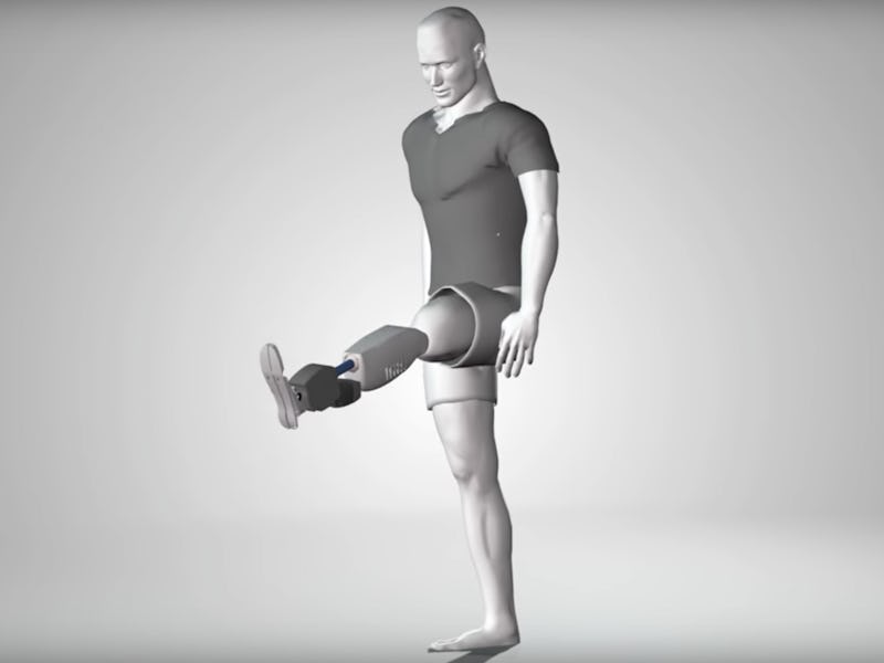
The agonist-antagonist myoplastic interface (AMI) consists of muscle pair constructs placed within the residual limb of an amputee.
The key comes from developing what the researchers call an agonist-antagonist myoneural interface. Basically, what that means is that they seek, through these muscle grafts, to rebuild the natural push-pull relationship of intact muscles. In an able-bodied person, each muscle system is counteracted by one that pulls in the opposite direction: flex your biceps and your triceps stretch, which not only prevents your body from straining too far and hurting itself but also gives you better control over your arm’s position in space. In existing prosthetics, this sense of control and self-awareness is missing, and people often have to keep an eye on what they’re doing with their artificial limbs.
Biophysicist Shriya Srinivasan says that she hopes this new focus on amputation and muscular reconstruction will improve the interdisciplinary efforts of amputation and prosthetic development that have only begun to emerge.
Abstract:
Prosthetic limb control is fundamentally constrained by the current amputation procedure. Since the U.S. Civil War, the external prosthesis has benefited from a pronounced level of innovation, but amputation technique has not significantly changed. During a standard amputation, nerves are transected without the reintroduction of proper neural targets, causing painful neuromas and rendering efferent recordings infeasible. Furthermore, the physiological agonist-antagonist muscle relationships are severed, precluding the generation of musculotendinous proprioception, an afferent feedback modality critical for joint stability, trajectory planning, and fine motor control. We establish an agonist-antagonist myoneural interface (AMI), a unique surgical paradigm for amputation. Regenerated free muscle grafts innervated with transected nerves are linked in agonist-antagonist relationships, emulating the dynamic interactions found within an intact limb. Using biomechanical, electrophysiological, and histological evaluations, we demonstrate a viable architecture for bidirectional signaling with transected motor nerves. Upon neural activation, the agonist muscle contracts, generating electromyographic signal. This contraction in the agonist creates a stretch in the mechanically linked antagonist muscle, producing afferent feedback, which is transmitted through its motor nerve. Histological results demonstrate regeneration and the presence of the spindle fibers responsible for afferent signal generation. These results suggest that the AMI will not only produce robust signals for the efferent control of an external prosthesis but also provide an amputee’s central nervous system with critical musculotendinous proprioception, offering the potential for an enhanced prosthetic controllability and sensation
