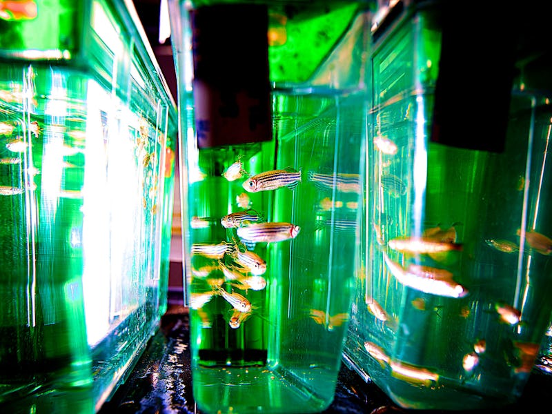
Zebrafish Melanoma Study Reveals Cancer's Mysterious Origins
Scientists have long wondered what sends potentially cancerous cells over the edge. Now they know.

After following a single fluorescent cell’s birth and spread through the body of a translucent fish, researchers at the Boston Children’s Hospital are now one step closer to answering a question that has long baffled scientists: Why don’t all cells with cancer genes become cancerous? In their study, which marks the first time a cancer cell has been visualized so early on in its lifetime, the researchers got close enough to the moment of a healthy cell’s betrayal to the cancerous dark side to figure out what exactly pushes it over the edge.
A perplexing issue in cancer research is the fact that scientists keep finding cells that express cancer genes but never become malignant. What the scientists have been doing is sort of like “cellular profiling,” so to speak: They use fairly visible markers — certain cancer-related genes, active or not — to guess whether a cell is potentially dangerous. But those genes, like a person’s clothes, aren’t enough to predict a cell’s fate. There’s something else — a trigger — that turns a somewhat-dangerous-looking cell into a bona fide cancer.
That trigger is what the team found. Ze’ev Ronai, Ph.D., a cancer specialist and scientific director at the Sanford-Burnham Medical Research Institute at La Jolla, called it “a significant advance in the field,” which is characteristically sedate scientist-speak for “this is a huge-ass deal.”
In a study published in the journal Science on Thursday, lead author Charles K. Kaufman, Ph.D. and his team revealed that to become truly cancerous, the skin cancer cells they followed in zebrafish required three things. The team already knew about the first two — a mutation in the gene BRAF (which also is found in human skin cancer) and the loss of the tumor suppressor gene p53 — but what their investigation revealed was the third factor, a change that causes the cell to revert back to the stem cell state.
In this case, it was the activation of a gene called Crestin, which seemed to give the newly induced “stem cell” the green light to go full cancer, activating the genes that cause it to proliferate. This growing clump of cells is what becomes the melanoma — a cancerous skin mole. Neighboring cells that also had the BRAF and p53 mutations but never got kicked back to the stem cell state didn’t become cancerous.
Conveniently, the same genes that get activated post-Crestin in fish tumors are the same ones that are turned on in humans. This is important for two reasons. It means melanoma formation is probably the same in fish and humans, and it also means that there’s probably a Crestin-like gene in human moles that could be spotted before it can trigger its cancerous cascade.
We’ve been trained to freak out when we find moles on our bodies. Are they cancerous? Are they just sunspots? So far, it’s been really hard to tell. Kaufman estimates that only one in tens to hundreds of millions of cells in a mole could become cancerous. But now that we know what cells look like as they’re about to go bad, it might become a lot easier to spot them — and deal with them — before they even begin to wreak havoc.